3D Dental Imaging Using In Dentistry
Oral Surgery
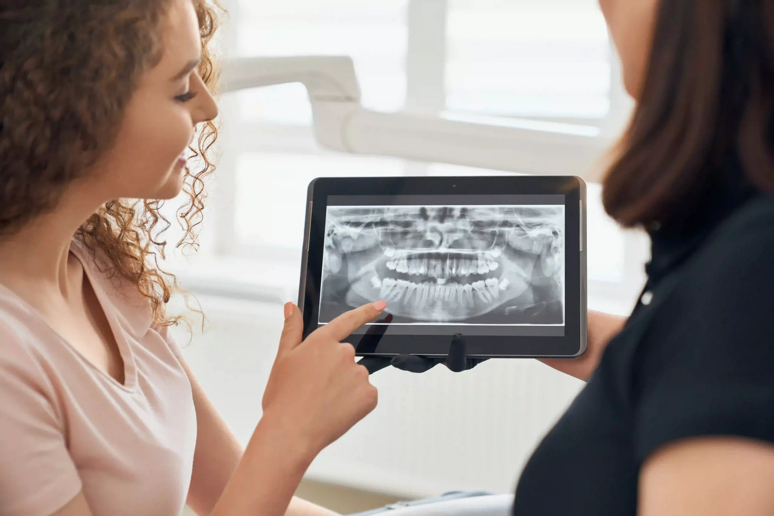
Table of Contents
The 3D Dental plays a major role in Dentistry. Imaging is equally important in the dental field as it is in the medical field. Since teeth are embedded within the jaw bone that is not visible to us, we need more complex aids to help us view and diagnose deeper internal areas of the human jaw. This is where radiography and CT scans come in.
While radiographs can give us a 2D Image of the bone, CT scans can give an even more accurate image in 3D.
Are you wondering what a 3D dental scanner is and how you can use it to improve your dental diagnosis and dental health? Here is everything that you need to know about a dental 3Dscanner:
What Is A 3D Dental Scanner?
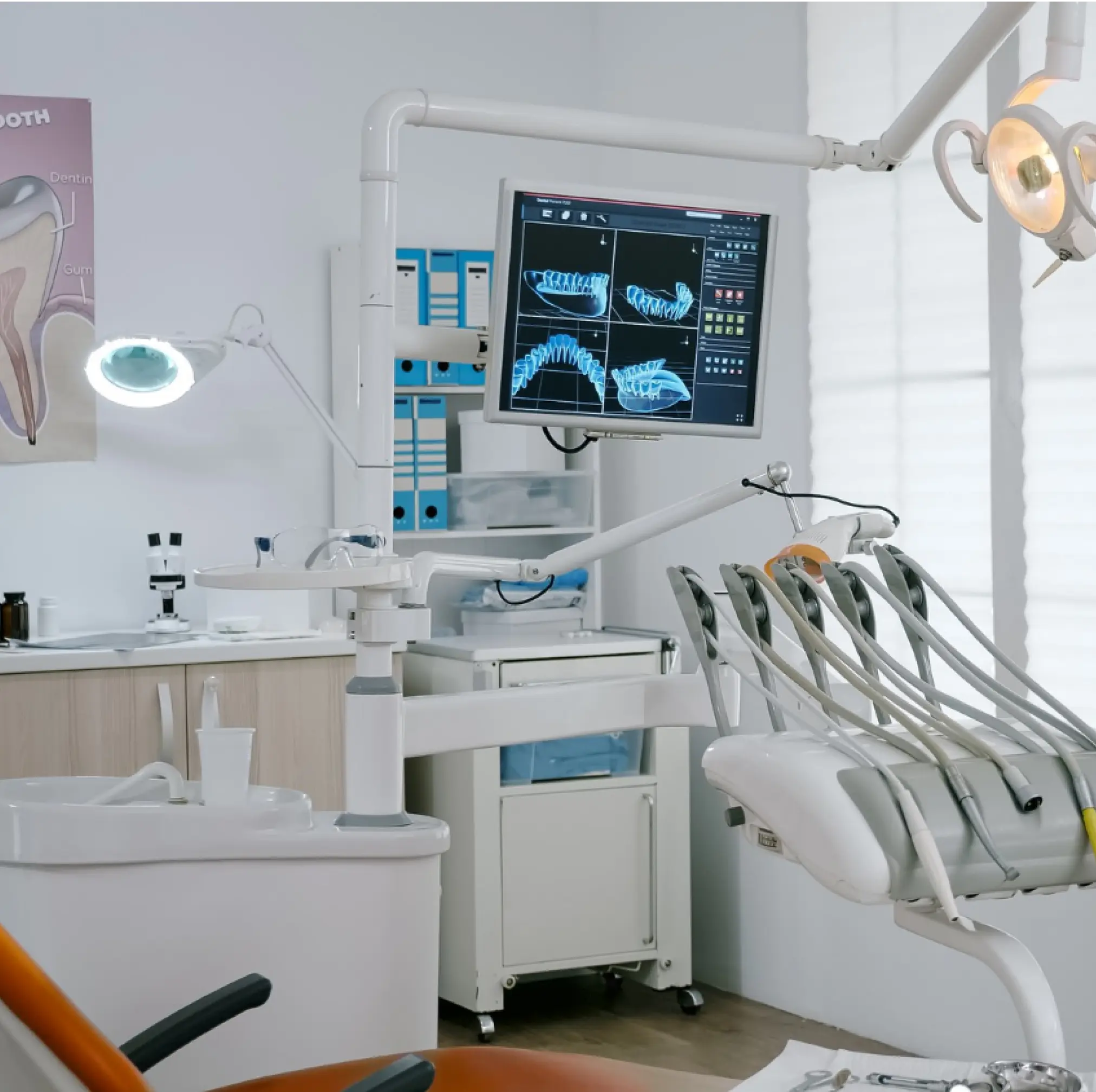
A CBCT or a 3D dental scanner takes images of internal structures of the bone and combines them to create a 3D image to allow healthcare providers to view them clearly and help with diagnosis. Unlike radiographs that only show the bone, the CBCT images allow dentists to view the soft tissues, airways, teeth, and bone surrounding it.
What Are The Uses Of A 3D Dental Scanner?
A CBCT dental scanner is the gold standard of imaging in dentistry and can help plan even better treatments for the following:
- Complicated wisdom tooth extractions
- Implant surgeries
- TMJ treatments
- Certain complicated extractions
- Root canal treatments
- Orthodontic treatment
The patient would be required to rest their chin on a chin rest in the CBCT scanner while it rotates around their head, shooting over 600 images of minimal radiation of the teeth, jaw and skull. The computer then combines these images to create a 3D image of the internal tissues, giving complete and clear images that trained technicians can read and understand.
What Are The Benefits Of Using A 3D dental imaging uses in dentistry?
There are many benefits of using a CBCT scanner; here are some of its benefits mentioned below:
Simple To Use
CBCT scanners are very easy to use. They are uncomplicated and always accurate. The patient must rest their chin while the machine rotates around their head once.
Minimal Radiation Exposure
While the scanner shoots up to 600 images, the radiation exposure is minimal and is even less than the radiation from X-rays. CBCT scanners are safer than X-rays.
Can View Soft Tissues Such As Blood Vessels And Nerves
CBCT scanners can also take images of soft tissues such as blood vessels and nerves, which was previously not possible to do with X-rays. Viewing nerves and blood vessels made it easier to plan complicated extractions.
Reduced Risk Of Nerve Injuries
Certain impacted wisdom teeth and multi-rooted teeth that are believed to lie close to the nerves are at risk of getting injured during extractions. With the help of CBCT imaging, nerve injuries can be avoided, and different angles can be used for extractions to avoid being too close to the nerve.
It Makes Implant Placement More Accurate
Implants require adequate bone quality and height for stabilization. CBCT scanners can be used to accurately assess bone quality and height to plan implant surgeries and increase their chances of success. The correct implant length can be selected without risking nerve damage, and the entire procedure can be clean and precise.
Reduces Risk Of Sinus Perforations
Upper teeth sometimes have very long roots and often curved ones. When extracting these teeth and placing implants, there is always a risk of damaging or perforating the maxillary sinus. Using a CBCT scanner, the bone height and the maxillary sinus can be seen in the images; therefore, there is minimal to no risk of perforating the maxillary sinus.
Painless And Quick
CBCT scanners are quick, efficient, and painless. They are easy to use and cause no pain or discomfort to the patient. X-rays can often be a little uncomfortable for patients and may often have to be repeated at different angles. This is not a problem when using CBCT scanners as they shoot upto 600 images from all possible angles to create the perfect 3D image.
Great For Orthodontic Treatment
CBCT scans are excellent for planning orthodontic treatment for teeth alignment. This is ideal for patients who wish to undergo orthodontic treatment. Virtual treatment plans can be made using the CBCT scanner for accurate and successful orthodontic treatments.
The VATECH Dental Scanner
Our clinic uses the latest Green X VATECH 4-in1 Digital X-ray system that encompasses Pano, Ceph {optional}, CBCT, and Model Scan. With VATECH’s extensive experience in the dental imaging field, the Green X provides high-quality Images with lower radiation by combining image processing, giving high-quality results. It increases precision in diagnostics and enhances treatment planning, providing the best care to your patients. It can take the following images:
- OPG or Panoramic images
- Cephalorams
- CBCT
- Model scan
The Final Word
At Dr. Paul’s Dental Clinic, we have the latest Digital Dental Scanner that can be used for dental imaging and the best treatment plans. Make an appointment today and meet with one of our trained dental professionals today. Our clinic employs the latest machines and materials to provide optimum treatment for all its patients and has the best skilled and trained professionals at its practice.
Also Read: Learn about the situations requiring wisdom tooth removal in our blog on Wisdom Tooth Extractions: When and Why Are They Required ?.
Book an Appointment With Your Doctor NOW!
Ready for a brighter smile? Schedule your appointment with Dr. Paul’s Dental Clinic today and experience exceptional dental care.
 Dr. Roy Thomas